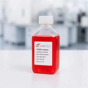
Product Details
- Product Name: PriGrow II Medium
- Product Type: Medium
- Catalog Number: TM002
- Unit Size: 500 mL
- Formulation: Serum-free and antibiotic-free basal medium
- Appearance: Liquid
- pH Range: 7.0-7.6
- Osmolarity: 264-292 mOsm/kgH₂O
- Endotoxin Level: ≤ 1 EU/mL
- Sterility: Pass
- Storage Condition: 2-8°C
- Shipping Condition: Shipped on ice packs
Overview
PriGrow II Medium is a specialized basal medium formulated for mammalian cell culture applications. Designed to provide optimal growth conditions, this medium offers flexibility for researchers by allowing the addition of specific growth factors, serum, or antibiotics as needed. Each lot undergoes stringent quality control testing to ensure batch-to-batch consistency in pH balance, sterility, and endotoxin levels.
This medium is suitable for a variety of cell culture applications, including primary cell expansion, immortalized cell line maintenance, and experimental research on cell differentiation and signaling pathways.
Key Features and Benefits
- Serum-Free and Antibiotic-Free: Provides a versatile foundation for cell culture, allowing customized supplementation.
- Optimized for Mammalian Cell Growth: Supports robust and consistent cell proliferation across various applications.
- Stringent Quality Control: Each lot is tested for sterility, osmolality, pH stability, and endotoxin levels to ensure a reproducible environment.
- Customizable Composition: Compatible with various growth factor and serum supplementation strategies.
- Validated for Performance: Designed to support adherence (if applicable), typical cell morphology, and metabolic activity in culture.
Key Ingredients and Special Additives
- L-Glutamin: 2.05 mM
- D-Glucose: : 11.1 mM
Quality Control (QC)
Each batch of PriGrow II Medium undergoes rigorous QC testing to maintain high reliability in cell culture applications. This includes:
- Cell Culture Performance Testing: Evaluation of adherence, morphology, and metabolic activity in tested cell lines.
- Sterility Testing: Ensuring an aseptic environment free from microbial or fungal contamination.
- Physicochemical Property Analysis: Monitoring of pH stability and osmolality.
- Endotoxin Screening: Ensuring endotoxin levels remain within safe limits for sensitive cell cultures.
Caution
This product is intended for research use only. It is not approved for therapeutic, diagnostic, or clinical applications.
Disclaimer
- This product is strictly intended for laboratory research purposes and should not be used in humans or veterinary applications.
- The manufacturer does not warrant the accuracy, completeness, or applicability of the product information beyond its intended research use.
- Any referenced literature or third-party citations are provided for informational purposes only and do not constitute an endorsement of specific results.
References
- Verma, Gaurav, Himanshi Bhatia, and Malabika Datta. “JNK1/2 regulates ER–mitochondrial Ca2+ cross-talk during IL-1β–mediated cell death in RINm5F and human primary β-cells.” Molecular biology of the cell 24.12 (2013): 2058-2071.
- Yong, Cai, et al. “The therapeutic effect of monocyte chemoattractant protein-1 delivered by an electrospun scaffold for hyperglycemia and nephrotic disorders.” International Journal of Nanomedicine (2014): 985-993.
- Kiratipaiboon, Chayanin, Parkpoom Tengamnuay, and Pithi Chanvorachote. “Glycyrrhizic acid attenuates stem cell-like phenotypes of human dermal papilla cells.” Phytomedicine 22.14 (2015): 1269-1278.
- Rubenstein, David A., et al. “Tobacco and e-cigarette products initiate Kupffer cell inflammatory responses.” Molecular immunology 67.2 (2015): 652-660.
- Galván, José A., et al. “Validation of COL11A1/procollagen 11A1 expression in TGF-β1-activated immortalised human mesenchymal cells and in stromal cells of human colon adenocarcinoma.” BMC cancer 14 (2014): 1-12.
- Levine, Corri B., et al. “Effects and synergy of feed ingredients on canine neoplastic cell proliferation.” BMC Veterinary Research 12 (2016): 1-11.
- Han, Zhezhu, et al. “Survivin silencing and TRAIL expression using oncolytic adenovirus increase anti-tumorigenic activity in gemcitabine-resistant pancreatic cancer cells.” Apoptosis 21 (2016): 351-364.
- Charan, Manish, et al. “GD2‐directed CAR‐T cells in combination with HGF‐targeted neutralizing antibody (AMG102) prevent primary tumor growth and metastasis in Ewing sarcoma.” International journal of cancer 146.11 (2020): 3184-3195.
- Han, Zhezhu, et al. “TGF-β downregulation-induced cancer cell death is finely regulated by the SAPK signaling cascade.” Experimental & Molecular Medicine 50.12 (2018): 1-19.
- Seoane, Marcos, et al. “Lineage-specific control of TFIIH by MITF determines transcriptional homeostasis and DNA repair.” Oncogene 38.19 (2019): 3616-3635.


