Product Details
- Catalog Number: T0843
- Unit Size: 1×10^6 cells / 1.0 ml
- Species: Human (Homo sapiens)
- Tissue: Peripheral blood
- Donor Information: CD34⁺-enriched mononuclear cells from a donor with aspirin-exacerbated respiratory disease
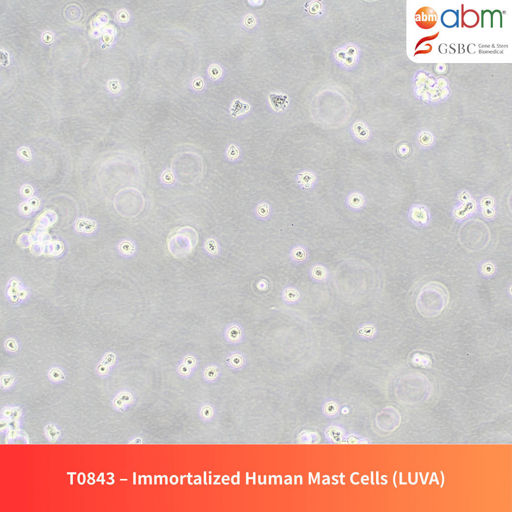
- Cell Type: Immortalized Mast Cells
- Morphology & Growth Properties: Suspension; round morphology (may attach in non-upright culture vessels)
- Biosafety Level: BSL-2
- Storage Conditions: Vapor phase of liquid nitrogen or below -130°C
- Shipping Conditions: Shipped on dry ice
- Format: Cryopreserved frozen cells
- Incubation Conditions: 37°C, 5% CO₂
- Immortalization Method: Spontaneous immortalization
Overview
LUVA cells are an immortalized mast cell line established from CD34⁺ progenitors cultured with stem cell factor (SCF), interleukin-6, and transient interleukin-3 exposure. These cells contain metachromatic cytoplasmic granules immunoreactive for tryptase, cathepsin G, and carboxypeptidase A3, and they express transcripts for FcεRI, c-kit, chymase, histidine decarboxylase, CPA3, and the type 1 receptor for cysteinyl leukotrienes.
LUVA can be propagated without SCF supplementation while maintaining functional c-kit and FcεRI signaling. Although FcεRI positivity is limited (~5% of cells), LUVA exhibits modest histamine release compared to LAD2, providing a reproducible system for mast cell biology and drug research applications.
Key Features and Benefits
- Defined Origin: Derived from CD34⁺ blood mononuclear cells of an aspirin-exacerbated respiratory disease donor.
- Granule Marker Profile: Positive for tryptase, cathepsin G, and carboxypeptidase A3.
- Functional Signaling: Retains c-kit and FcεRI signaling pathways.
- SCF-Independent Growth: Cultured without exogenous stem cell factor.
- Selective and Reliable Model: Limited FcεRI expression with modest histamine release, offering a stable and reproducible system for immunology and drug research.
Culture & Handling Guidelines
Recommended Culture Conditions
- Culture Vessel: SpheroWell™ T75 Flask (G7543) or upright PriCoat™ T25 Flasks (G299)
- Growth Medium: PriGrow X Series Medium (TM0843) supplemented with:
- 2 mM L-glutamine (G275)
- 1% Penicillin/Streptomycin Solution (G255)
- Incubation: Maintain at 37°C in a humidified atmosphere with 5% CO₂.
- Seeding Density: 1 × 10⁵ – 3 × 10⁵ cells/ml.
- Doubling Time: 24-48 hours.
- Note: Do not culture or freeze with FBS.
Thawing Protocol
- Thaw cells quickly in a 37°C water bath (≤ 2 minutes), keeping the vial cap above water.
- Decontaminate the vial exterior with 70% ethanol before transferring into a biosafety cabinet.
- Transfer cell suspension into a 15 mL sterile tube with 5 mL pre-warmed complete medium.
- Centrifuge at 125×g for 5–7 minutes and discard the supernatant.
- Resuspend the pellet in fresh complete medium and dispense into a T75 culture flask.
Subculturing Guidelines
- Change the medium every 2–3 days and keep cell density ≤ 1 × 10⁶ cells/mL.
- Split cultures at a 1:2 to 1:20 ratio (1:10 typically reaches confluence in 3–4 days).
- Centrifuge at 125×g for 5 minutes, resuspend in fresh medium, and transfer into new culture vessels.
Cryopreservation Guidelines
- Cryopreservation Medium: Complete medium supplemented with 8% DMSO.
- Freezing Procedure: Freeze gradually at -1°C/min before transfer to liquid nitrogen.
Related Products
- Recombinant Human SCF (E. coli) – Z100815
- Recombinant Human TGF Beta-1 (TGFB1) – Z101555
- Recombinant Human TNF – Z101385
Disclaimer
- This product is for research use only. Not intended for human or animal diagnostic, therapeutic, or clinical applications.
- The user is responsible for verifying the suitability of this product for their specific experimental conditions.
- Handling and usage should comply with all relevant biosafety regulations.
- No warranties, express or implied, are provided regarding the performance of this product outside of the recommended conditions.
References
- Kirshenbaum, Arnold S., et al. “Demonstration that human mast cells arise from a progenitor cell population that is CD34+, c-kit+, and expresses aminopeptidase N (CD13).” Blood, The Journal of the American Society of Hematology 94.7 (1999): 2333-2342.
- Kirshenbaum, Arnold S., et al. “Characterization of novel stem cell factor responsive human mast cell lines LAD 1 and 2 established from a patient with mast cell sarcoma/leukemia; activation following aggregation of FcεRI or FcγRI.” Leukemia research 27.8 (2003): 677-682.
- Laidlaw, Tanya M., et al. “Characterization of a novel human mast cell line that responds to stem cell factor and expresses functional FcεRI.” Journal of allergy and clinical immunology 127.3 (2011): 815-822.